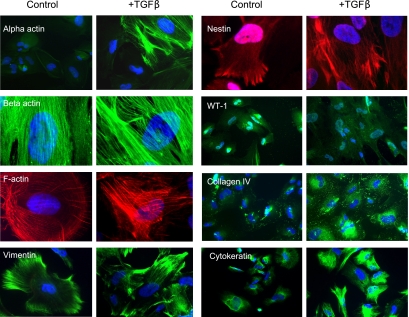
FIG. 2.
Changes in the expression of key markers of differentiation in immortalized human podocytes in response to treatment with TGF-β1 (10 ng/mL) for 3 days when compared with control cells. Immunofluorescence staining for α-actin, β-actin, F-actin, vimentin, nestin, WT1, collagen IV α3, and cytokeratin, with a blue nuclear counterstain (DAPI). (A high-quality color representation of this figure is available in the online issue.)

