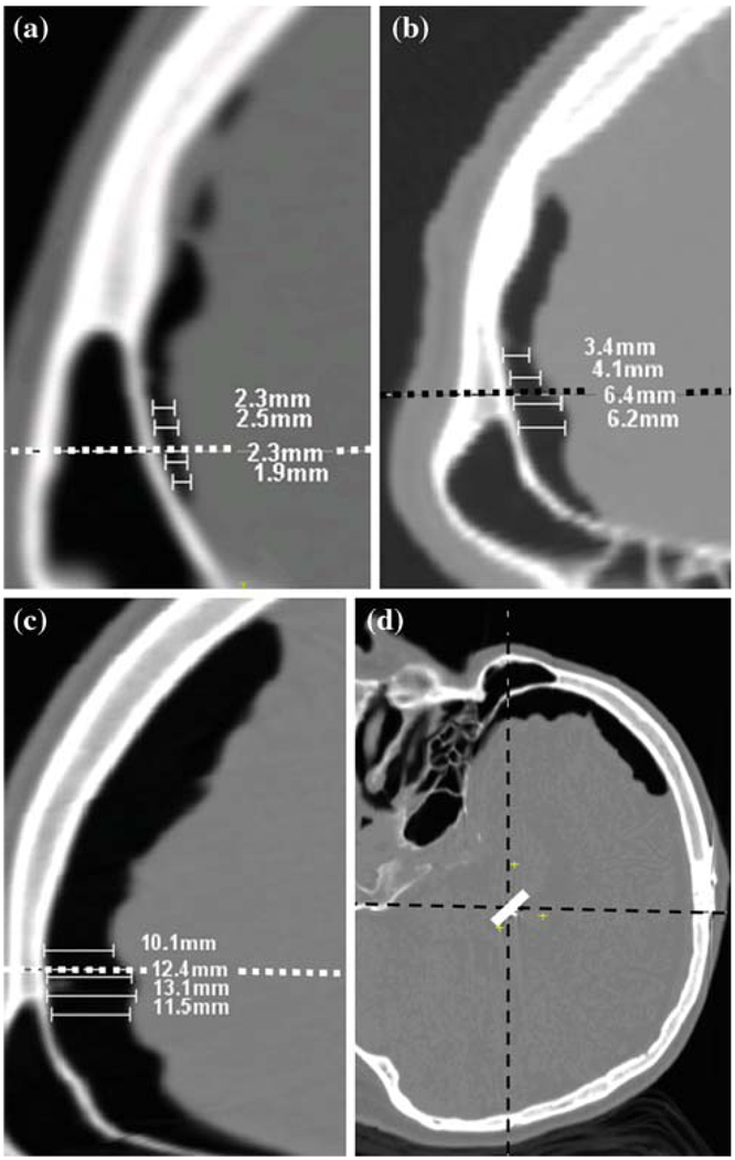
Fig. 1.
Frontal portion of a sagittal slice (after a 90° rotation for easy visualization) on post-operative CT for patients in a low, b medium, c large shift groups showing air pocket. Width of the air pocket is measured from the inner table of the calvarium at the lead level as shown. Sample measurements in mm are shown. d Typical head orientation in the scanner with the patient lying supine. The dashed horizontal line in a, b and c is a vector passing through the implant in the direction of gravity

