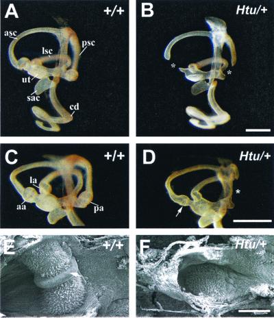
Figure 1.
Paintfilled inner ears at E16.5 showing missing or smaller ampullae in Htu/+ mutants. (A–D) Medial views of control and Htu/+ ears. (B) The phenotype where both ampullae are absent. (D) The phenotype in which just the posterior is missing. Asterisks in B and D indicate the positions of the missing ampullae. Note the smaller anterior ampulla in the Htu/+ mutant (arrow in D). (E and F) Scanning electron microscopy of the anterior crista in the control and mutant at P3. Note the small, flat appearance and the missing eminentia cruciata (arrow in E) in the mutant crista (F). aa, anterior ampulla; asc, anterior semicular canal; cd, cochlear duct; la, lateral ampulla; lsc, lateral semicircular canal; pa, posterior ampulla; psc, posterior semicircular canal; ut, utricle. (Scale bar in B = 500 μm for A and B, in D = 500 μm for C and D, and in F = 100 μm for E and F.)

