Figure 2.
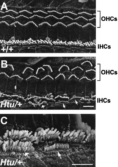
Scanning electron micrographs of the organ of Corti in Htu/+ mice demonstrate neuroepithelial patterning defects within this organ. (A and B) Low-power views from the basal turn of the organ of Corti of a control (A) and heterozygote (B), showing the reduced number of outer hair cell rows (OHCs) in the mutant. Anomalies within the inner hair cell (IHC) row can also be observed in B and C (higher power), including extra inner hair cells (fat arrow) and atypical hair cells (thin arrows). (Scale bar in B = 5 μm for A and B. Scale bar in C = 5 μm.)

