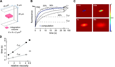
Figure 1.
Diffusion measured by fluorescence recovery after photobleaching with confocal detection. A) Bleach geometry (rectangular volume 6×18×20 μm) and signal integration volume for 1-layer diffusion. B) Photobleaching recovery for 20-μm-thick layers of PBS containing 2 mg/ml FITC-dextran (70 kDa) with indicted sucrose to increase viscosity. Bleach depth was 25–35% (shown normalized for comparison). Fluorescence was detected in a z plane at the center of the 20-μm-thick solution layer and represents integrated signal in a detection volume of 6 μm × 6 μm × 2 μm. Blue circles represent theoretical fluorescence recovery kinetics for the 20% sucrose solution computed using the finite-element method (number of elements, 272667; relative tolerance, 10−6). C) Pseudocolored concentration profiles of relative concentration at t = 0, 0.5, 1, 20 s for diffusion coefficient 4 × 10⁻⁸ cm2/s. D) Fitted t1/4 and t1/2 for photobleaching measurements as in B (means ± se, n=5).

