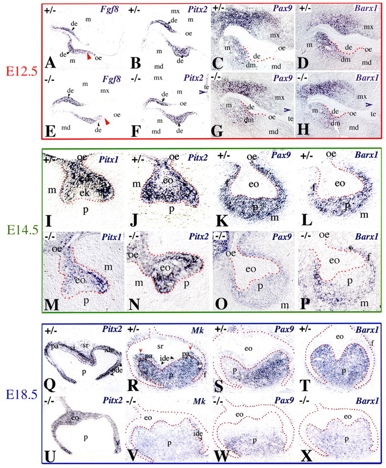
Fig. 7.

Expression of Fgf8, Pitx1, Pitx2, Pax9, Barx1 and Mk in molars of Jag2+/− and Jag2−/− mouse embryos. In situ hybridisation on frontal cryosections of E12.5 (A-H), E14.5 (I-P) and E18.5 (Q-X) embryos. Red dotted lines indicate borders between dental epithelium (de) and mesenchyme (dm). (A-H) Fgf8 (A,E), Pitx2 (B,F), Pax9 (C,G) and Barx1 (D,H) expression. Fgf8 expression is slightly restricted in Jag2−/− dental epithelium (red arrowheads in A and E). (I-P) Pitx1 (I,M), Pitx2 (J,N), Pax9 (K,O) and Barx1 (L,P) expression. A weak Pax9 signal is seen in dental papilla (p) and follicle (f) and downregulation of Barx1 expression is seen in dental papilla of Jag2−/− teeth. (Q-X) Pitx2 (Q,U), Mk (R,V), Pax9 (S,W) and Barx1 (T,X) expression. Pitx2 expression in inner dental epithelium (ide), stratum intermedium (si) and outer dental epithelium (ode) in Jag2+/− teeth (Q). Downregulation of Pitx2 in preameloblasts (pa) and stellate reticulum (sr). In Jag2−/− embryos (U), Pitx2 is expressed in all epithelial cells. Downregulation of Mk (V), Pax9 (W) and Barx1 (X) expression in dental papilla and follicle of Jag2−/− embryos. Mk transcripts are absent in preameloblasts of Jag2−/− teeth (V), but are present in Jag2+/− teeth (areas indicated by red arrowheads in R). eo, enamel organ; ek, enamel knot; m, mesenchyme; md, mandible; mx, maxilla; oe, oral epithelium; te, tongue epithelium.
