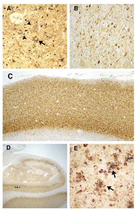
FIGURE 1.

Silver stain and tau pathology in proband (A). Bielschowsky silver stained sections from the proband demonstrate significant neurofibrillary tangle (arrowheads) and neuritic plaque (arrow) formation in the frontal cortex. Note the coreless plaque (arrow). Tau immunohistochemistry revealed severe immunopositive staining of neurofibrillary pathology in the frontal cortex (B and C, PHF1 antibody) and hippocampus (D, PHF-1 antibody). (E) Double immunolabeling of plaquelike structures in the hippocampus with prion protein (3F4, purple) and tau (PHF-1, brown) antibodies revealed a close anatomic relationship between these pathologic changes (arrows). Original magnification, A and B, ×63; C, ×13; D, ×7; E, ×126.
