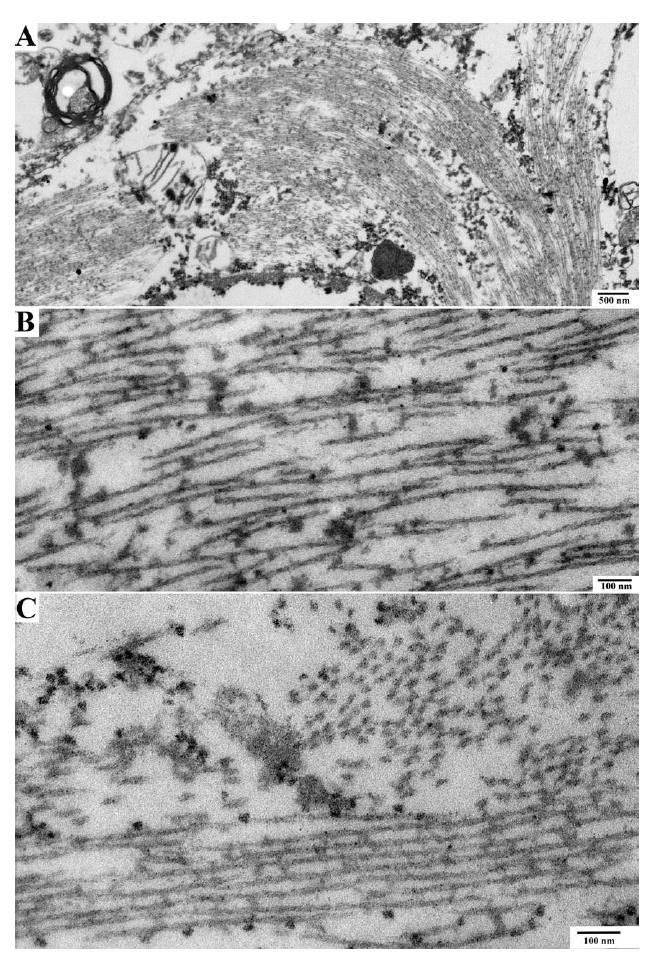
FIGURE 3.

Electron microscopy of neurofibrillary tangles in proband. Electron microscopic pictures of neuronal cytoplasm show the presence of neurofibrillary tangles. (A) Cytoplasm of a nerve cell containing a neurofibrillary tangle. (B, C) High-power image of neurofibrillary tangle shows paired helical filaments seen longitudinally (lower portion of the image) and in cross-section (upper right portion of image).
