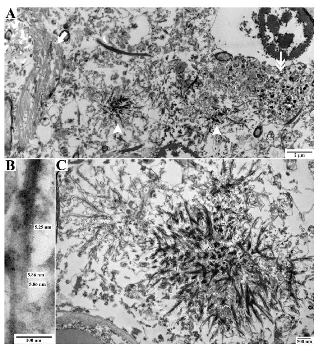
FIGURE 4.

Electron microscopy of prion protein (PrP) deposition in proband. Electron microscopic pictures of neuropathologic lesions in the neuropil. (A) The neuropil shows a neurofibrillary tangle on the left (arrow), PrP amyloid deposits in the center (arrowheads), and a dystrophic neurite on the right (double arrow). (B) High-power image of filaments from a PrP amyloid plaque. (C) Starlike PrP amyloid plaques.
