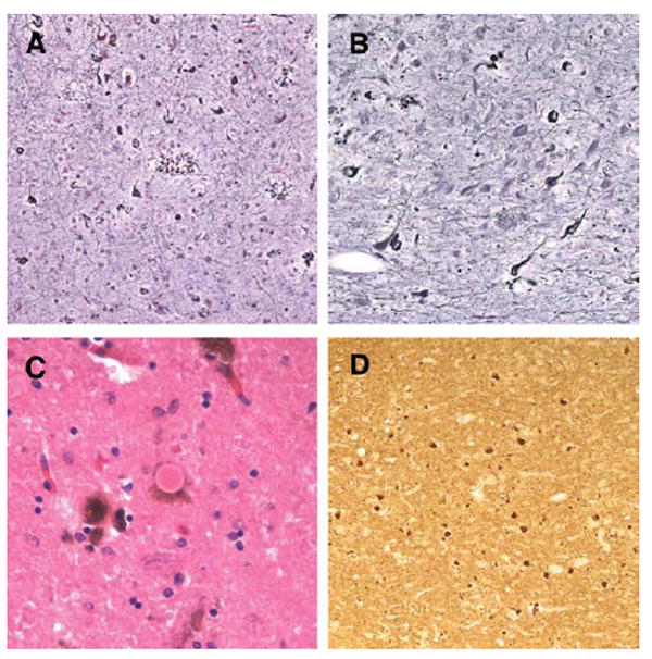
FIGURE 5.

Histologic pathology in the proband’s mother. Holmes silver staining revealed severe neurofibrillary tangles and neuritic plaque pathology in the mother’s frontal cortex (A) and hippocampus (B). In addition, both classic Lewy bodies (C) and alpha-synuclein immunopositive inclusions and neurites (D) were observed. Original magnification, A and B, ×63; C, ×126; D, ×32.
