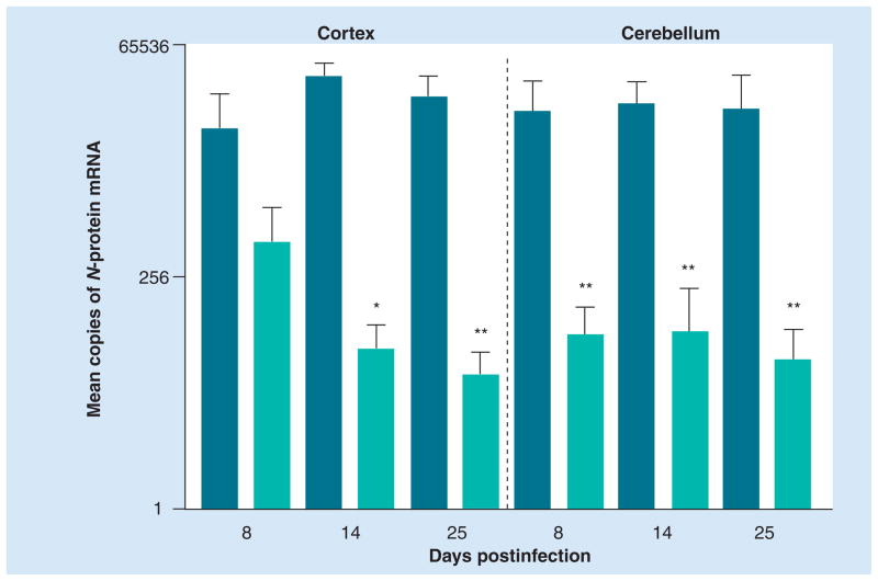
Figure 1. Passive administration of rabies virus immune sera from normal mice inhibits CVS-F3 replication in the CNS tissues of B-cell-deficient mice.
Groups of five JHD−/− mice were infected with 105 focus-forming units of CVS-F3 intranasally. A total of 5 days later, mice were injected intraperitoneally with 0.5 ml of either normal or rabies virus immune serum. At the time points indicated, cortex and cerebellar tissues were collected and virus replication was assessed by real-time quantitative RT-PCR. Virus replication is expressed as the mean plus the standard error of the mean copies of rabies virus nucleoprotein mRNA in 50 ng of cDNA isolated from CNS tissues. Statistically significant differences in gene expression between tissues from mice receiving normal and rabies virus immune serum, determined by the Mann–Whitney test, are denoted by *(p < 0.05) and **(p < 0.01).

