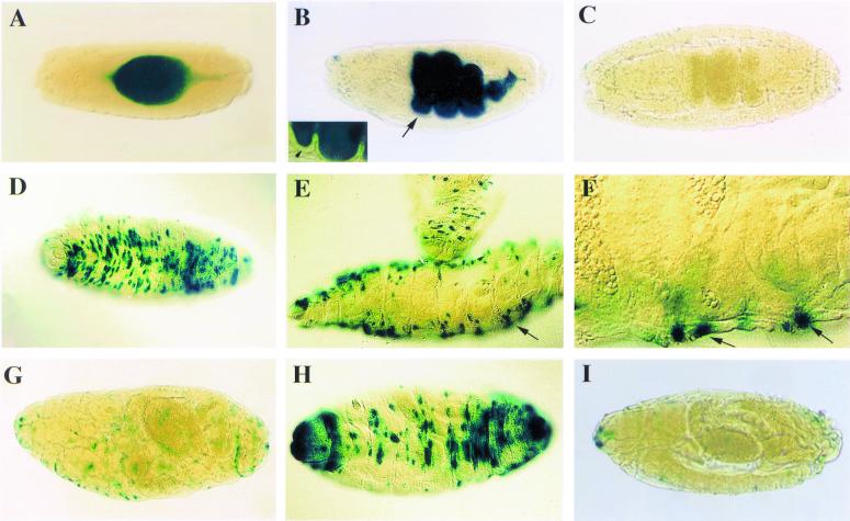
Figure 3.
Bacterial infection induces CecA1-lacZ expression in embryos. Anterior is to the left, and dorsal is up in lateral views. (A–G) Induction of β-gal expression in the yolk and epidermis of A10 transgenic embryos after injection of LPS, bacteria, or PBS 3–4 h before fixation and staining. (A) Dorsal view of a stage-14 embryo injected with E. cloacae. (B) Ventrolateral view of a stage-16 embryo injected with LPS. The staining is strong throughout the yolk present within the developing midgut and hindgut. The expression is within the yolk sac but excluded from midgut epithelium (arrows and Inset). (C) Lateral view of a stage-16 embryo injected with PBS. (D) Dorsal view of a stage-17 embryo injected with LPS around 12 h AEL. Expression no longer can be induced in the yolk region, but staining is established in epidermal cells throughout the embryo. The staining appears as an irregular pattern of transversal rows including both groups of cells as well as individual cells. (E) Lateral view of a stage-17 embryo injected with LPS demonstrating the uneven distribution of stained cells in the embryos with a concentration of positive cells on the dorsal and ventral sides. (F) Magnification of the same embryo as in E with a slightly different focus shows that the stained cells lie just in the embryo surface (arrows). (G) β-gal staining in a stage-17 A10 CecA1-lacZ embryo after injection of sterile PBS. (H–I) Induction of β-gal expression in the epidermis of embryos at stage 17 3–4 h after LPS injection carrying different CecA1-lacZ constructs. (H) Dorsal view of an A12 embryo showing the same pattern of β-gal staining as in A10 embryos. (I) Lateral view of an A15 embryo displaying no inducible expression. The stages of embryonic development are according to ref. 38.

