Figure 4.
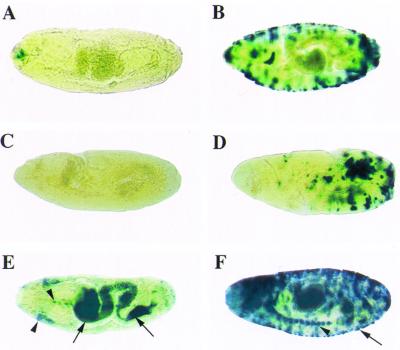
Analysis of the requirement of GATA-binding factors for inducible CecA1 expression in the yolk and epidermis, respectively. Anterior is to the left, and all embryos are in lateral view with dorsal up, except for E which is in ventral view. Embryos were injected with LPS before (A, C, and E) or 12 h after (B, D, and F) egg laying to induce CecA1 expression in the yolk or in the epidermis, respectively. (A–B) β-gal staining in A16 CecA1-lacZ embryos with a mutated GATA site. No expression is observed in the yolk (A), whereas expression in the epidermis is strong (B). (C–F) β-gal expression in A10 CecA1-lacZ embryos in srp6G mutant background. No CecA1 expression could be detected in the yolk of homozygous srp6G embryos (C) but is evident clearly in individual cells and in patches of cells in the epidermis (D). The development of the srp6G embryos (C and D) is arrested, and the embryos do not complete germ-band retraction and dorsal closure as their heterozygous siblings (E and F). In embryos heterozygous for srp6G and for the ftz-lacZ blue balancer, the β-gal staining is evident both from the CecA1 promoter (arrows) in the yolk (E and F), in the epidermis (F), and from the ftz promoter (arrowheads) in a weak stripe in the anterior part of the embryo (E) and in the ventral nerve chord (E and F; refs. 23 and 24).

