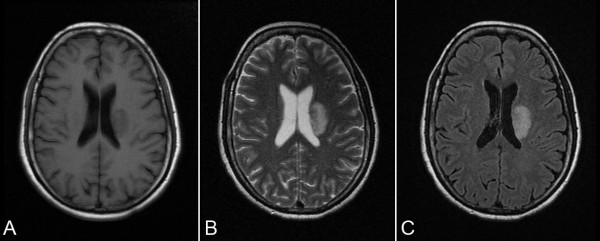
Figure 2.
T1-FLAIR, T2-weighted and T2-FLAIR images from a patient show focal infarct at left corona radiate. (A) T1-FLAIR obtained 2 days after stroke show a hypointense area at left corona radiate. (B) T2-weighted image and (C) T2-FLAIR images obtained at W1 show a hyperintense area at left corona radiate.

