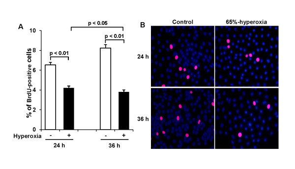
Figure 3.
Effect of 65%-hyperoxia on fetal type II cell proliferation. (A) Graphical depiction showing BrdU-positive cells in hyperoxic and normoxic cells. The results are represented as the mean ± SD from 3 different experiments. (B) Representative fluorescence immunocytochemistry fields of E19 type II cells exposed to 65%-hyperoxia for 24 h and 36 h and parallel control samples. BrdU positive cells are labeled red. Nuclei were counterstained with DAPI (blue). Scale bar = 50 μm.

