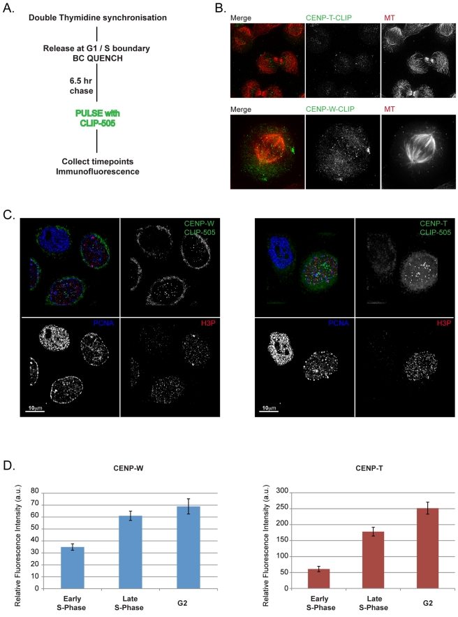
Figure 3. CENPs -T and -W assemble predominantly in late S-phase and G2.
(A) Schematic description of the CLIP quench-chase-pulse-chase experiment used to assay the timing of assembly of CLIP tagged CENPs -T and -W at centromeres using synchronized HeLa cells. (B) CLIP-tagged CENPs -T and -W are localized at centromeres prior to the onset of anaphase, indicating that newly synthesized CENP-T and CENP-W assemble at the centromere in the proximal cell cycle. (C) Progressive assembly of pulsed CENP-T and CENP-W in S-phase and G2. Cells were labelled with PCNA and phospho-histone H3 antibodies to document position in the cell cycle. Cells judged to be in earlier stages of S-phase have no detectable CLIP signal at centromeres, while cells later in S-phase and G2 have robust centromere-associated CLIP signal. (D) Centromere-associated CENP-T-CLIP and CENP-W-CLIP fluorescence were quantified relative to progression through the cell cycle, showing an increased signal intensity at centromeres coinciding with progression through S-phase and G2.

