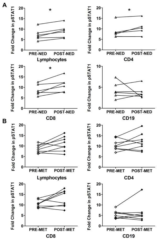
Figure 3.
Correlation of IFN-α induced pSTAT1 and long-term clinical outcome in HDI patients. At the time of clinical follow-up, patients were classified as exhibiting no evidence of disease (NED) or metastases (MET). The IFN-α induced fold change of pSTAT1 in melanoma patients' lymphocytes and lymphocyte subsets were assessed in patients exhibiting A) NED and, B) MET before (pre-) and after (post-) the HDI induction phase. Two-sided paired Wilcoxon-Mann-Whitney tests were performed on melanoma patients pre- and corresponding post- PBMCs and adjusted P-values < 0.05 were considered significant. CVs were calculated by dividing the standard deviation with the mean of the fold changes multiplied by 100. (*Lymphocytes: p = 0.042, 95% CI: -3.12 to -0.25, CV: pre-NED 37.4%, post-NED 34%; *CD4: p = 0.042, 95% CI: -4.77 to -0.48, CV: pre-NED 39.6%, post-NED 36.9%; *CD8: p = 0.042, 95% CI: -5.12 to -0.13, CV: pre-NED 30.2%, post-NED 29.9%).

