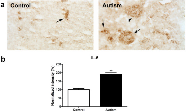
Figure 1.
IL-6 expression increased in the cerebellum of autistic subjects. Immunohistochemistry studies were carried out on cerebellar homogenates from 6 autistic subjects and 6 age-matched controls using an IL-6 antibody (dilution 1:100). Stronger immunostaining of IL-6 (dark brown color indicated by an arrow) was present in the autistic samples (a). Immunostaining density was quantified using Image J analysis (b). Data are shown as mean ± SEM.

