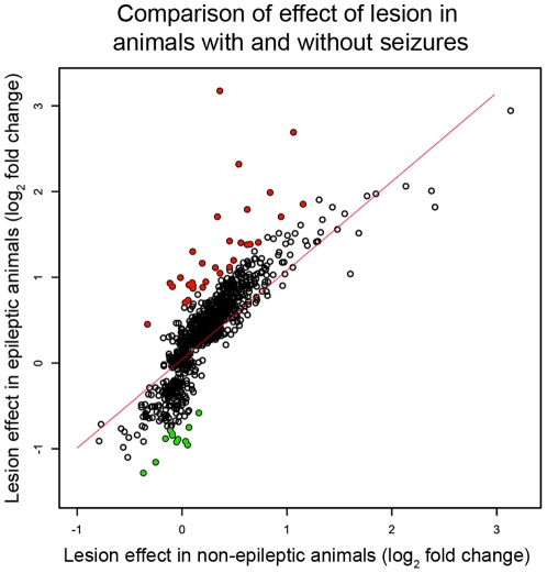
Figure 4. Most differentially expressed genes within the epileptogenic region were related to the effects of the lesion.
We examined the effect of the lesion on the genes that were specifically differentially expressed within the epileptogenic region to determine whether they were plausible candidate genes to be associated with epileptogenicity. The scatter plot depicts the comparison of the lesion-induced fold changes between the animals with and without seizures for all of the genes that were specifically differentially expressed in the epileptogenic region. The x-axis represents the mean fold changes between the lesioned and non-lesioned hippocampi in animals without seizures and the y-axis represents the means fold changes between the lesioned and non-lesioned hippocampi in animals with seizures. All of the fold changes were transformed using log2. We found that there was a high correlation between the expression changes in animals with seizures and animals without seizures (r = 0.84), demonstrating many of these expression changes were related to the lesion. To remove this confounding factor, we used a linear regression to define the relationship between the effect of the lesion in animals with seizures and the effect in animals without seizures (red line). We then removed all genes that were within two standard deviations of this line (empty points), and after subtracting these genes, we identified 40 genes whose gene expression changes were not related to the lesion or seizures (12 down-regulated – green; 28 upregulated – red).

