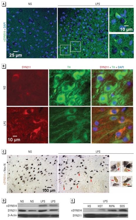
Figure 3.
Nitration of aggregated α-syn and formation of cytoplasmic inclusions in Tg mice 5 months after LPS injection. (A) Nitrated human α-syn (labeled with nSYN514 antibody) formed aggregates and was mainly perinuclear in the SN of LPS-injected Tg mice (center), whereas nSYN514 staining in NS-injected Tg mice (left) was scant. Magnifications (right) are from areas shown in the LPS section. (B) The SN of LPS-injected Tg mice contained human α-syn aggregates (red) in TH-IR neurons (green). (C) Cytoplasmic inclusions containing nitrated human α-syn (brown) in nigral neurons (with nuclei stained in dark blue) indicated by red arrows in LPS-injected Tg mice (center) and magnified photomicrographs (right) from the LPS section. The SN of NS-injected Tg mice (left) did not exhibit obvious staining for nitrated α-syn. (D) Western blotting shows nitrated human α-syn in midbrain extracts from LPS-injected Tg mice but not NS-injected Tg mice. (E) The accumulation of nitrated human α-syn was observed primarily in RIPA and SDS fractions from midbrains of Tg mice. At least four mice in each group were used for immunostaining and immunoblotting. For magnifications in C, bars = 10 μm.

