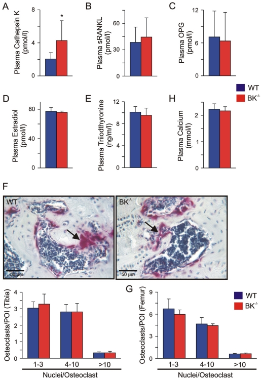
Figure 2. Enhanced plasma Cathepsin K, but normal endocrinology and osteoclastogenesis in juvenile BK−/− mice.
(A) Enhanced plasma Cathepsin K levels in juvenile BK−/− mice. Statistics derived from 9 WT and 8 BK−/− mice. (B, C) Osteoblast-derived factors influencing osteoclast activity were not altered in juvenile BK−/− mice. Statistical summary of plasma sRANKL (B) and OPG (C) from WT and BK−/− mice (n = 6–12 per genotype). (D, E) Endocrine factors regulating osteoclast activity were not altered in juvenile BK−/− mice. Statistics of plasma estradiol (D) and plasma triiodothyronine (E) from WT (n = 12–17) and BK−/− mice (n = 12–16). (F) Upper: Apparent number of osteoclasts in native BK−/− proximal tibia differed not when compared to WT. Nuclei of TRAP5b-positive (purple) osteoclasts (arrow) were stained with hematoxylin (blue). Lower: Statistical summary of osteoclast quantification in tibia. (G) Statistics of osteoclast quantification in distal femur. In the cortical and cancellous compartment of the evaluated bones, TRAP5b-positive cells (osteoclasts) were counted by two independent persons in a double-blinded manner in five non-overlapping pictures of interest in each of 8–10 consecutive slices with an inter-slice-interval of 120 µm. The tibiae and femurs were derived from 4 WT and 4 BK−/− mice. (H) Statistics of plasma Calcium in WT and BK−/− mice (n = 9 per genotype). All data are means±SD; *P<0.05.

