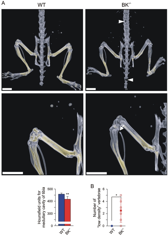
Figure 4. Reduced bone mineral density (BMD) in juvenile BK−/− mice.
(A) Upper: Representative dorsal views of volume rendering of legs, pelvic bone and backbone from a juvenile WT (left) and a BK−/− mouse (right), and a magnification of their leg (Middle). BMD within the bones is reflected by a yellow-colour-scaling, whereas “yellow” encodes for high BMD values. Note the black gap (arrow) in BK−/− fibula, and the “low-density” BK−/− vertebrae and bones of the mouse tail (triangle), which was due to the very low BMD within that bone region; bar: 5 mm. Lower: Statistics of Hounsfield units in medullary cavity of tibia (n = 4–6 per genotype). (B) Statistical summary of “low-density” vertebrae apparent only in BK−/− mice (n = 4–6 per genotype). All data are means±SD; *P<0.05; **P<0.01.

