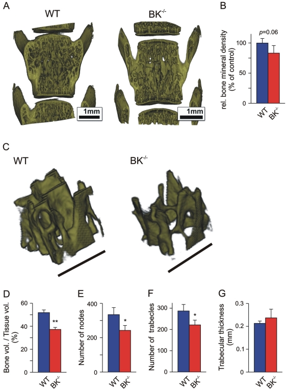
Figure 6. Increased porosity of BK−/− vertebral trabecular meshwork.
(A, B) Representative 3D reconstructions of the 4th lumbar vertebra obtained by high-resolution µCT (A), and corresponding statistics of relative bone mineral density (B) from juvenile WT and BK−/− mice (n = 4). (C–F) Representative reconstructions of a cubic region of interest (0.5×0.5×0.5 mm3) within the trabecular meshwork of the 4th lumbar vertebra (C), and corresponding statistics of bone volume (BV)/tissue volume (TV)-ratio (D), number of nodes (E), trabecles (F) and trabecular thickness (G), evaluated in three cubic regions of interest (0.5×0.5×0.5 mm3). Bars: 1 mm (A), 500 µm (C); all data are means±SD; *P<0.05; **P<0.01.

