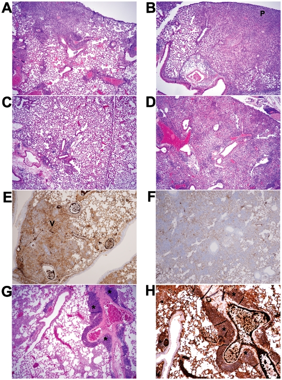
Figure 4. Intranasal infection with C. muridarum induces more extensive inflammatory changes in the lungs of TLR2-deficient mice compared to wild type mice.
C57BL/6 or TLR2 KO mice on the same background were intranasally inoculated with PBS (mock) or 5×10³ IFU/mouse of C. muridarum Nigg. At the indicated time points, mice were euthanized and lungs were removed for tissue processing, as described in the Methods. A–D and G show routine H&E staining; P = dense PMN infiltrate, shown as deep purple-staining cells. E–F and H show vimentin and CD19 staining by immunohistochemistry, respectively. (A) C57BL/6, day 7; (B) TLR2-deficient, day 7; (C) C57BL/6, day 14; (D), TLR2-deficient, day 14. (E–F) Lungs from infected TLR2-deficient mice (F) fail to stain for vimentin (V) at 5 days post infection compared to C57BL/6 mice (E). (G–H) Lungs from infected TLR2-deficient mice show evidence of iBALT (*) at 7 days post infection when stained with H&E (G) or anti CD19 Ab to detect CD19-expressing B cells (H). Original magnification 40×. This figure is representative of three independent experiments performed.

