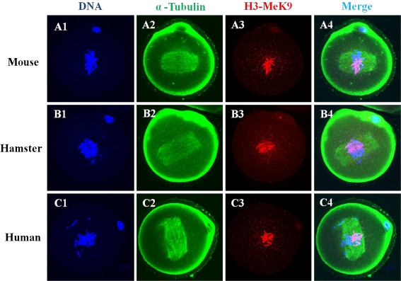
Fig. 5.
Laser scanning confocal microscopic images of microtubules, H3K9 methylation and chromatin are shown at the metaphase stage of mouse zygotes after ICSI with mouse, hamster and human sperm. Metaphase chromatin from mouse oocytes after ICSI with mouse (A1-A4), hamster (B1-B4) and human (C1-C4) sperm was seen in half of the chromatin stained with H3-MeK9. All images are labeled for microtubules (green), DNA (blue) and H3-MeK9 (red)

