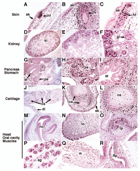
Figure 1.
FoxM1 expression in the mouse embryo. Paraffin sections were prepared from mouse wild type embryos harvested at E13.5 (G–I and R), E15.5 (A, B, D and J–P) or E18.5 (C, E, F and Q). Sections were used for immunohistochemistry with FoxM1 antibodies (brown) and counterstained with nuclear fast red (red nuclei). Nuclear FoxM1 staining was detected in hair follicles (A and C), follicles of vibrissae (B) and keratinocytes of basal layers (A–C). FoxM1 was also observed in developing kidney (D–F), cartilage (J–L), epithelial and mesenchymal cells of pancreas and stomach (G and I). FoxM1 expressing cells were detected in nasal cavity (M), tongue (N), tooth primordium (O), salivary glands (P), muscles (Q) and the Rathke's pouch (R). sk, skin; hf, hair follicle; fv, follicle of vibrissae; ki, kidney; gl, glomeruli; li, liver; pa, pancreas; st, stomach; ca, cartilage; di, diaphragm; nc, nasal cavity; to, tongue; tp, tooth primordium; sg, salivary gland; m, muscle; Rp, Rathke's pouch. Magnification: (D, G, J and M) x50; (C, F and L) x400; (A, E and N–P) x100; remaining parts, x200.

