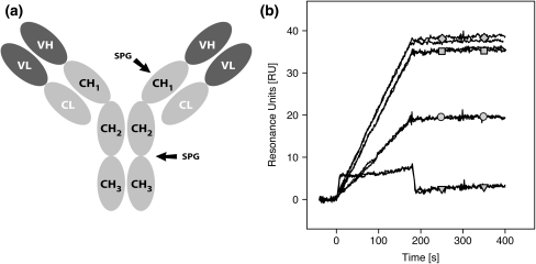
Fig. 7.
Mapping of Zmab25 binding site by competitive binding analyses. Possible binding sites on mouse IgG1 for the Zmab25 variant were mapped using a competitive binding analysis employing an immunoglobulin binding protein with known binding sites. a Schematic figure showing an IgG antibody protein with its different regions and the binding sites for SPG in Fc and Fab, respectively, indicated. b Biosensor sensorgrams resulting from duplicate injections of 10 nM mAb3 alone (gray diamonds); mAb3 in a 100 nM (gray squares), 1 μM (gray circles), or 10 μM (gray triangles) solution of the recombinant two-domain protein G construct (C2–C3) over a sensor chip surface containing the Zmab25 variant

