Fig. 1.
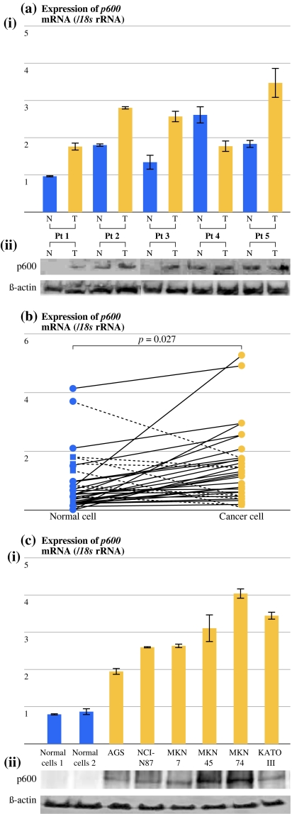
Gastric cancer cells express high levels of p600 mRNA. a Expression levels of p600 mRNA and protein in gastric cancer tissues and normal gastric tissues. (i) Reverse transcriptase–polymerase chain reaction (RT-PCR). (ii) Western blot test. β-Actin was used as the internal control for immunoblotting. The expression level of p600 protein was consistent with the expression level of p600 mRNA, and there was a tendency for gastric cancer tissues to have higher expression of p600 than normal gastric tissues. b Expression levels of p600 mRNA in laser-microdissected gastric cancer cells and normal gastric mucosal cells was determined by quantitative RT-PCR (n = 42). Gastric cancer cells showed significantly higher expression levels of p600 mRNA than normal gastric mucosal cells (P = 0.027). c Expression levels of p600 mRNA and protein in gastric cancer cell lines and normal gastric mucosal cells obtained from surgically resected normal gastric mucosa. (i) RT-PCR. (ii) Western blot test. β-Actin was used as the internal control for immunoblotting. Gastric cancer cell lines showed higher levels of p600 expression than normal gastric mucosal cells

