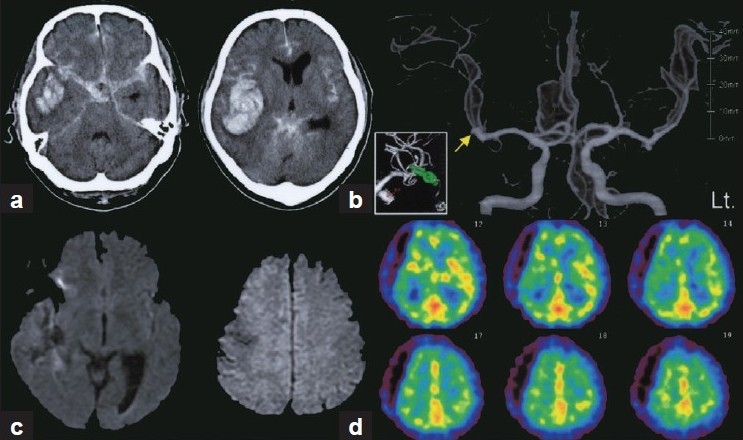
Figure 1.

(a) CT scans showing SAH combined with a massive right temporal intracerebral hematoma. (b) Preoperative 3-dimensional CT angiography showing bilateral MCA aneurysms. Ruptured aneurysm on the right side (arrow) was successfully clipped (inset). Clinical deterioration attributable to vasospasm was suspected based on findings of no apparent ischemic lesion on diffusion-weighted MR images (c) and relatively decreased rCBF in the left ACA and MCA territories on Tc-99 m HMPAO SPECT (d)
