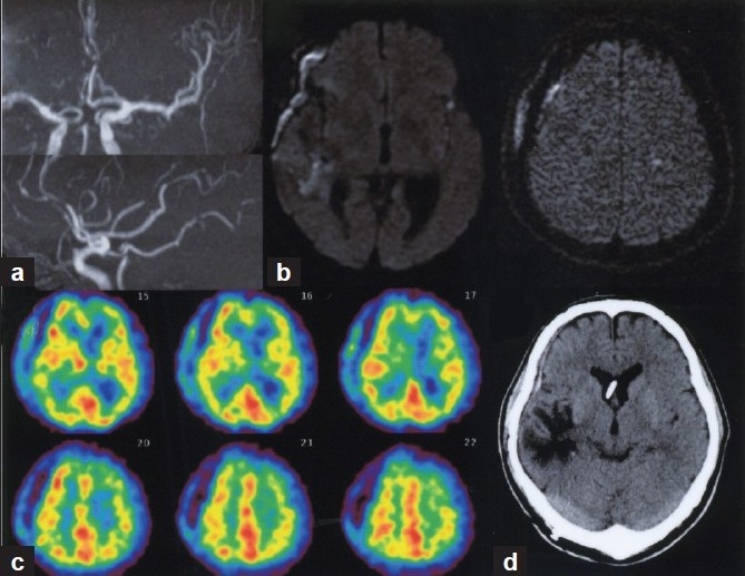
Figure 4.

(a) Follow-up MR angiography on day 14 showing improvement of vasospasm. (b) Diffusion-weighted MR images demonstrated no additional ischemic findings after repeated endovascular therapy. (c) Tc-99 m HMPAO SPECT confirmed normalized rCBF distribution in the left hemisphere. (d) CT scan at 2 months after SAH showing no apparent ischemia in the left hemisphere
