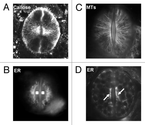
Figure 3.
(A) Callose immunolabeling in a differentiating stoma of A. nidus. Confocal-laser-scanning-microscope image constructed from seven consecutive optical paradermal sections. The callose fibrils are radially arranged around the stomatal pore. x850 (B) MT immunolabeling in a closed A. nidus stoma. The MTs underneath the periclinal walls are radially arranged around the stomatal pore (cf. A). x1,000. (C and D) Immunolabeling of HDEL ER proteins in a differentiating (C) and a mature closed stoma (D) of A. nidus. In the stoma shown in (C) distinct ER aggregations are localized at the margins of the wall thickenings (squares in C) deposited at the stomatal pore region. The arrows in (D) point to local ER aggregations in the cytoplasm adjacent to the walls at the stomatal pore region. C: x1,000; D: x750.

