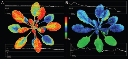
Figure 1.
Partial exposure to excess light and induction of SAAR is associated with systemic gradient-like changes of foliar NPQ and changes in PEPS. Arabidopsis thaliana Col-0 rosettes were grown at low-light conditions (LL, 100 µmol photons m−2s−1) and were partially exposed to excess light (EL, 2,000 µmol photons m−2s−1). (A) Left, controls that were LL grown with no excess light exposure. (B) Right, the same rosette partially exposed to EL for 60 min (three bottom leaves with the lowest NPQ value). NPQ estimated the nonphotochemical quenching from Fm to F′m is monitoring the apparent heat losses from PSII and is calculated from (Fm/F′m) − 1, where Fm is maximal chlorophyll fluorescence of dark-adapted PSII (all QA molecules are reduced), F′m = is maximal chlorophyll fluorescence of light-adapted PSII (all QA molecules involved in photosynthetic electron transport from water in provided light condition are reduced). Changes in PEPS (mV) were measured in bundle sheath cells of EL exposed leaves and in leaves undergoing SAAR. One bundle sheath cell of directly exposed leaf and another in a leaf undergoing SAAR were measured during whole experiment (60 min with 15 min periods of light on and off). Strong plasma membrane electrical potential changes were observed directly after switching on and off EL. Pattern of PEPS changes in directly EL exposed leaves and leaves undergoing SAAR in LL are symmetric. When depolarization is observed in EL exposed leaves, hyperpolarization is induced in systemic leaves. Other experimental data are presented in Szechynska-Hebda and colleagues.17

