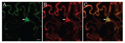
Figure 1.
Nucleocytoplasmic localization of VBF. GFP-tagged VBF was transiently coexpressed with free DsRed2 in Nicotiana benthamiana epidermis following microbombardment. (A) GFP-VBF. (B) DsRed2. (C) Merged image. GFP signal is in green, DsRed2 signal is in red and overlapping GFP and DsRed2 signals are in yellow. Arrow indicates the cell nucleus. All images are single confocal sections. Bar = 10 µm.

