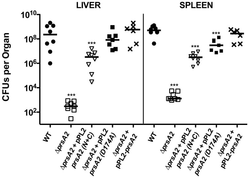
Figure 5. The PPIase domain is critical for restoration of virulence in animal models of infection.
Mice were intravenously infected with 2 × 10⁴ CFU of wild type (●), ΔprsA2 (□),ΔprsA2 + pPL2- prsA2 (X), ΔprsA2 + pPL2-prsA2 N+C (▽), and ΔprsA2 + pPL2-prsA2 D174A (■). At 72 hours post-infection livers and spleens were isolated, homogenized, and plated for bacterial CFU on solid BHI agar plates. CFU from the livers and spleens of a minimum of five mice are shown as scatter plots. Solid lines indicated the median value for each group. Statistical significance was calculated using a 1-way ANOVA with Tukey’s multiple comparison test. Statistically significant difference compared to WT and ΔprsA2 + pPL2- prsA2 are indicated (***, P < .0001).

