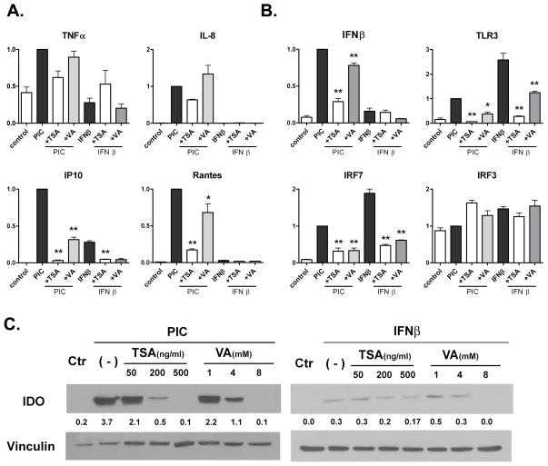
Figure 4. The effect of HDAC inhibitors on astrocyte inflammatory and innate immune gene expression.
Primary human fetal astrocytes were pretreated with TSA at 400 mg/ml or VA at 4 mM for 1h, and then stimulated with PIC at 20 μg/ml or IFNβ 10 ng/ml for an additional 6 h. Q-PCR was performed as described in Figure 2 and Figure 3 legend. Data are shown for cytokines and chemokines (A) and molecules involved in TLR3 signaling (B). Pooled, normalized (PIC = 1) data from two separate astrocyte cases are shown. Mean ± SEM. ANOVA with Dunnett’s post: * = p <0.05, ** = p < 0.01. (C) Western blot analysis of astrocytes for IDO protein expression. Astrocytes were treated with varying concentrations of TSA or VA as indicated for 1 h, then with IFNβ or PIC for 24h. The numbers are densitometric ratios to vinculin, a protein loading control. The data are representative of two separate experiments with similar results.

