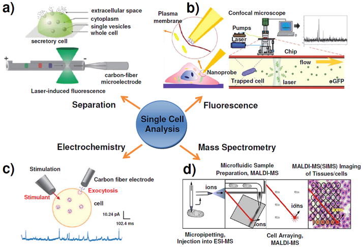
Figure 1.
Diagram showing the four major approaches to chemical analysis in, at, and of single cells, with an emphasis on exocytosis measurements. (a) Single cell analysis of exocytosis with capillary electrophoretic separation, which is capable of selectively sampling from localized regions within a single secretory cell (upper panel); Two of the most popular separation-based single cell detection schemes are LIF detection and electrochemical detection (lower panel). Reproduced from reference 51 with permission. (b) Fluorescence detection of extracellular lactate by an optical fiber based nanobiosensor (left panel); the general setup for the real time fluorescence analysis of single secreted proteins from a single yeast cell which is trapped in the chip by an electrical field is shown; enhanced green fluorescence protein molecules in the cell secretes can be detected by confocal microscopy (right panel). Reproduced from reference 60 and reference 282 with permission. (c) The typical setup for amperometry and typical amperometric data of a single cell in which exocytosis is stimulated by a pipette containing a stimulant. Reproduced from reference 90 with permission. (d) Schematics of the four mass spectrometry-based approaches for single cell metabolomics. Reproduced from reference 170 with permission.

