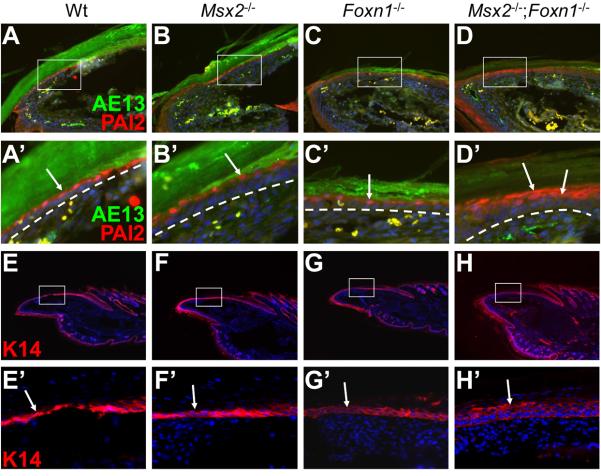
Figure 4. Hyperplastic nail bed in Msx2−/−;Foxn1−/− double mutant mice.
In addition to the keratogenous zone, PAI2 is also expressed in the suprabasal layer of the nail bed epithelium (A, A′, arrow). In Msx2 and Foxn1 single mutant nails, only one layer of cells in the nail bed is labeled, similar to wild type (B, B′, C, C′, arrows). However, PAI2 is detected in 2 to 3 layers of suprabasal cells in the double mutant nail bed (D, D′, arrows). AE13 staining marks the overlying nail plate (A-D, A′-D′). K14 is normally expressed in the nail matrix and the nail bed including both basal and suprabasal layers. In wild type, Msx2 and Foxn1 single mutant nail bed, K14 antibody stains two layers of cells. (E-G, E′-G′, arrows). However, more than three layers of cells are stained in the double mutant nail bed (H, H′, arrow). A′-D′ and E′-H′ are higher magnifications of boxes in A-D and E-H, respectively. White dashed lines demarcate the border between the nail bed and the underlying mesenchyme. Digit 4 of the hind limbs is used for assay. In all panels, the nail tips are orientated towards the left side.

