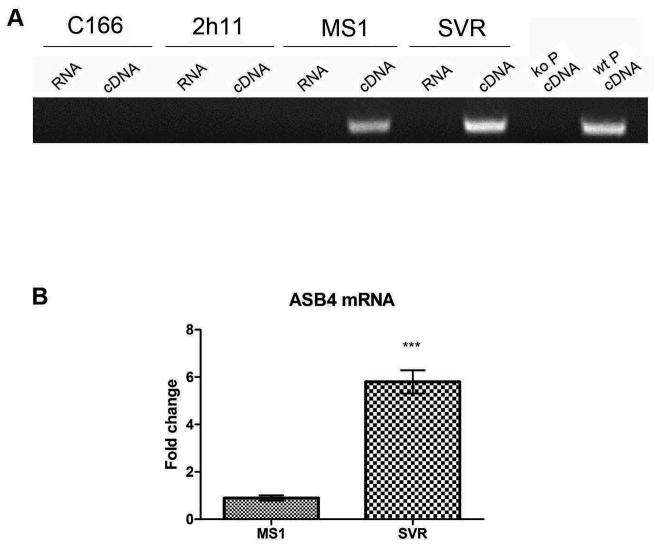
Figure 1.
Expression of ASB4 in endothelial cells. (A) C166, 2h11, MS1 and SVR cells were cultured to confluency, RNA was purified and RT-PCR with primers for ASB4 performed. RNA that had not been reverse transcribed was used as a control for genomic contamination. Placental cells from ASB4 knockout (ko P) and wild-type (wt P) mice were used as negative and positive controls for ASB4 expression. (B) Real time PCR analysis was performed on untreated MS1 and SVR cells and ASB4 expression normalized to MS1 cells. Results are shown as mean ± SD of three independent experiments. ***p < 0.001.

