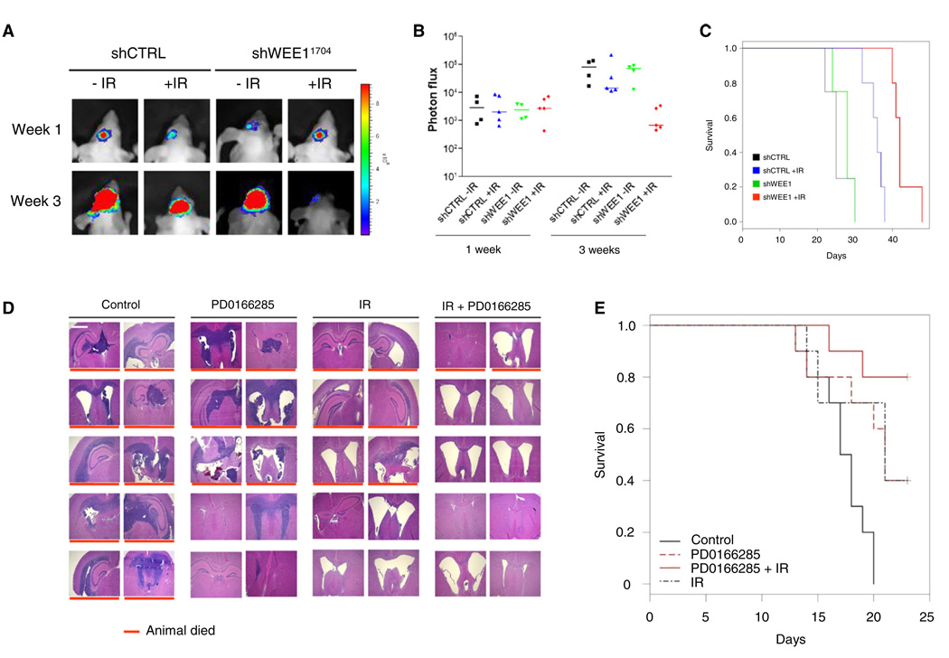
Figure 7. In Vivo Analysis of WEE1 Inhibition in an Invasive E98 GBM Mouse Model.
(A) E98-FM cells stably expression shWEE1 or shControl were grown in the brains of nude mice. The mice were divided in two arms of which one was irradiated with 3.5 Gy. Representative bioluminescence imaging results are shown 7 days and 3 weeks after onset of tumor growth.
(B) Relative photon flux at 7 days and 3 weeks after onset of tumor growth. (n = 4/group for control and n = 5 for IR treated).
(C) Survival curves of the mice in (B).
(D) E98 GBMs were grown in the brain of nude mice, irradiated with 8 Gy, and injected intraperitoneally with 500 µl of 20 µM of PD0166285 or PBS control for 5 consecutive days. Depicted are hematoxylin and eosin staining of irradiated E98 GBM tumors treated with PD0166285 or PBS control. Red lines indicate nonsurvivors. Size bar represents 1000 µm.
(E) PD0166285 enhances radiosensitivity in vivo. Depicted is survival (n = 10/group), log rank p = 0.001.
See also Figure S5.

