Fig. 1.
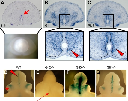
Expression pattern of hedgehog signaling genes in the male GT at E15.5. A, Shh is expressed in the UPE (arrow in the cross-section). B and C, Gli1 (B) and Ptc1 (C), representative hedgehog-responsive genes, are expressed in the mesenchyme adjacent to the UPE (arrowheads in the highly magnified pictures). Dotted lines indicate the boundary between the epithelium and mesenchyme of the developing GT. D–G, The LacZ staining pattern of the del5-LacZ line with several Gli mutant allelic backgrounds at E15.5. In the wild-type mouse embryos (WT), the LacZ signals are detected in the mesenchymal region adjacent to the UPE (D, arrowhead). An asterisk indicates the preputial gland. The Gli2 KO embryos exhibit a hypoplasic GT and groove-like structure in the ventral GT (arrow) without LacZ signals derived from the del5-LacZ allele (E). The ablation of Gli3 leads to enhanced LacZ signals, although lacking overt morphological GT defects (F). The Gli1 mutant GT does not show apparent GT phenotypes, and the LacZ signal is detected almost unaltered (G). No sexually dimorphic expression pattern is observed, and the representative images from male embryos are shown.

