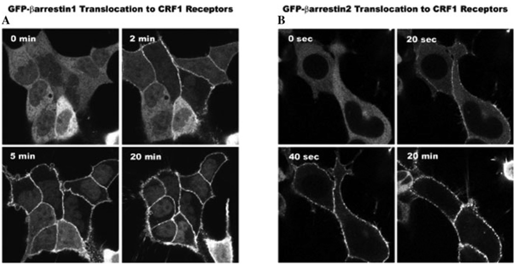
Figure 3.
Recruitment of β-arrestins by the agonist-activated CRF1 receptor. Confocal images depict the translocation of cytosolic β-arrestin1-GFP (A) or β-arrestin2-GFP (B) to membrane CRF1 receptors recombinantly expressed in HEK293 cells. A slower redistribution of β-arrestin1 to the cell surface occurred after (2-, 5-, 20-min) treatment with 200 nM CRF while a dramatically more rapid and robust recruitment of β-arrestin2 developed in cells after (20 sec, 40 sec, 20 min) treatment with 200 nM CRF.

