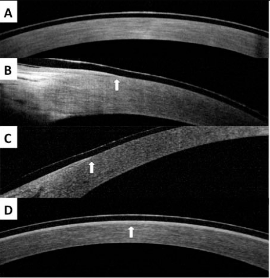
Fig. (1).
SD-OCT images from patient in case report. A, First post-operative day over central cornea vertex, showing absence of epithelial layer under TCL. B, First post-operative day over nasal limbus, showing edge of epithelial layer (arrow). C, First post-operative day over temporal limbus, showing edge of epithelial layer (arrow). D, Third post-operative day, showing complete epithelial layer under TCL (arrow).

