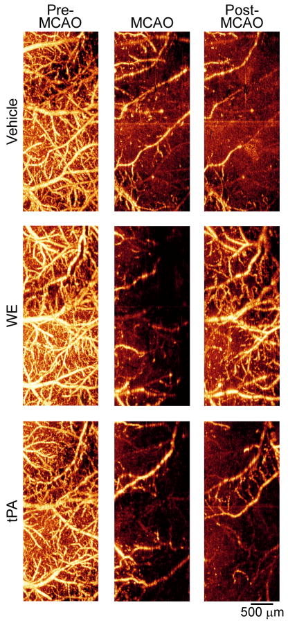
Figure 1. Optical microangiography (OMAG) of the cerebral cortex verifies extensive cortical hypoperfusion during and after MCAO.
Transcranial non-invasive OMAG was used to image the brains of mice through intact skull before MCAO, during MCAO, and post-MCAO. Bright areas indicate perfused vessels (moving blood) and dark regions indicate absence or reduction of perfusion.

