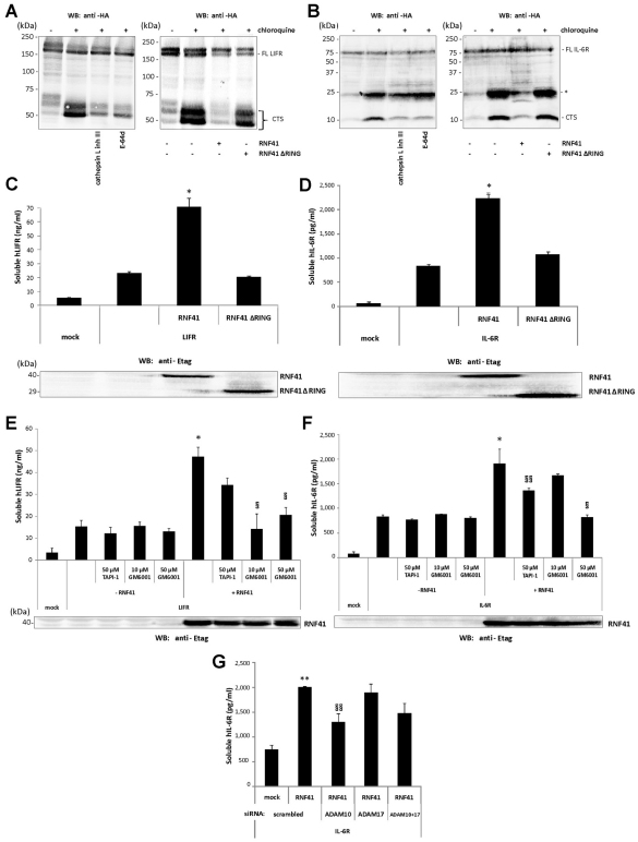
Fig. 6.
RNF41 blocks LIFRα and IL-6Rα cleavage by cathepsin L and enhances their shedding. (A,B) Chloroquine stabilises C-terminal LIFR and IL-6Rα fragments, which are blocked by cathepsin L inhibition (left panels) or RNF41 expression (right panels). HEK293T cells transiently transfected with C-terminal HA-tagged human LIFRα (LIFRα–HA) (A) or IL-6Rα (IL-6Rα–HA) (B) were left untreated (DMSO) or were incubated overnight solely with chloroquine or together with the indicated protease inhibitors (left panels); or were cotransfected with a full-length Etag–RNF41 or Etag–RNF41 ΔRING construct and left untreated or incubated with chloroquine (right). Cell lysates were analysed by western blotting (WB). FL, full length; *, intermediate IL-6Rα cleavage product; CTS, C-terminal stub. (C,D) RNF41 enhances LIFRα (C) and IL-6Rα (D) shedding. Cell media supernatants from the transfectants in the right panels of Fig. 6A,B were analysed for soluble LIFRα and IL-6Rα levels. E-tagged RNF41 or RNF41 ΔRING expression was verified by western blotting. (E,F) RNF41-enhanced LIFRα (E) and IL-6Rα (F) shedding is reversed by metalloprotease inhibitors. HEK293T cells transfected with hLIFRα–HA or hIL-6Rα–HA with or without full-length Etag–RNF41 were incubated overnight in starvation medium with TAPI-1 or GM6001 and soluble receptor levels in the cell medium supernatants were quantified. E-tagged RNF41 expression was verified by western blotting. (G) RNF41-enhanced IL-6R shedding is reversed by ADAM10 silencing. HEK293T cells, reverse transfected with siRNA targeting ADAM10 or ADAM17, were cotransfected the next day with hIL-6Rα–HA with or without full-length Etag–RNF41 and soluble IL-6Rα levels in the cell medium supernatants were quantified. Values are means ± s.d. (n=3). *P<0.01 or **P<0.05 (Student's t-test) compared with – RNF41; §P<0.01 or §§P<0.05 (Student's t-test) compared with +RNF41.

