
Figure 3.
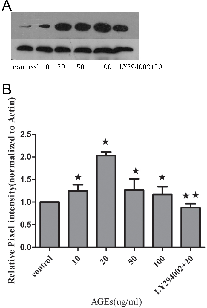
Protein expression of Robo1 in human retinal pigment epithelium cells was measured by immunoblotting with normalization to β-actin expression in retinal pigment epithelium cells. A: A representative photograph of the immunoblot analysis for Robo1 expression in human retinal pigment epithelium (RPE) cells. B: Relative Robo1 protein levels between the control group, advanced glycation end-products (AGEs) group, and LY294002-treated group. Values provided are mean±SD of three independent experiments. Asterisks denote values significantly different between the treated group and control group (p<0.05). Double asterisks denote values significantly different between the LY294002 (10 uM) group and AGEs (20 ug/ml) group (p<0.01).The pixel intensity of the control group was set to 1.