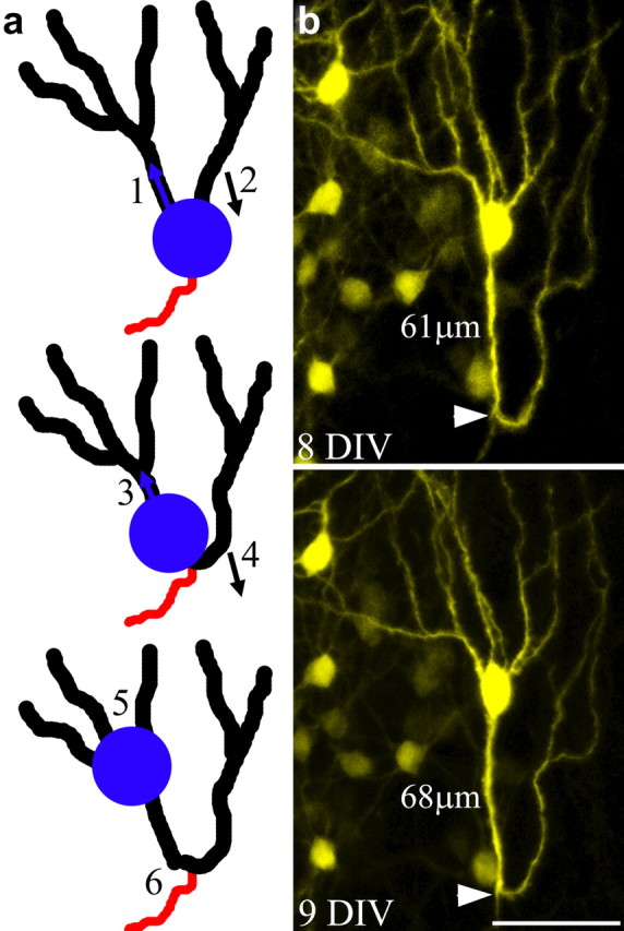
Figure 4.

Model depicting granule cell dysmorphogenesis following somatic translocation into an apical dendrite. a, As the soma moves into the initial segment of the left apical dendrite (1), the right apical dendrite is “left behind,” gradually shifting its position from the apical to the basal pole of the cell (2). Continuation of this process leads to a shortening of the initial dendritic segment on the left (3), and conversion of the right apical dendrite into a recurrent basal dendrite (4). In extreme cases, further migration of the soma leads to the absorption of branch points, converting these structures into primary dendrites (5), and the formation of dendritic loops projecting toward the hilus (6). b, An example of a granule cell exhibiting a recurrent basal dendrite with a pronounced dendritic loop and an unusually large number of apical dendrites, the putative end product of the processes outlined in a. Note that the axon (white arrowhead) projects off the base of the dendritic loop, suggesting that this was the original location of the cell body. Scale bar, 50 μm.
