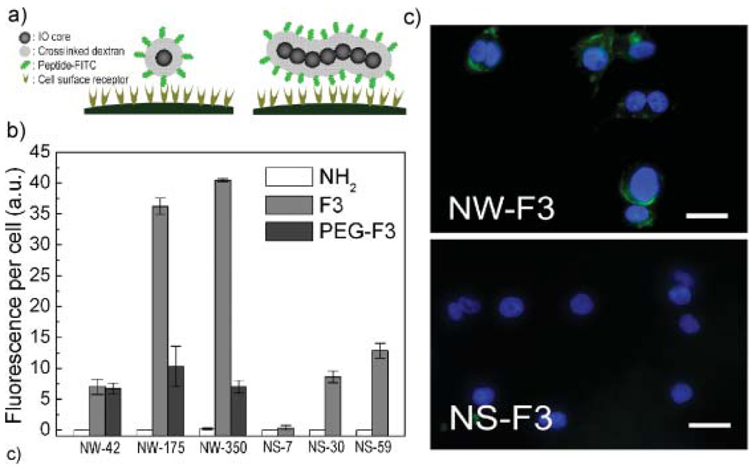
Figure 2.
Internalization of nanoworms (NWs) and nanospheres (NSs) conjugated with F3 peptides into MDA-MB-435 cells. a) Conceptual scheme illustrating the increased multivalent interactions expected between receptors on a cell surface and targeting ligands on a NW compared with a NS. b) Fluorescence data comparing the efficiency of cellular internalization for various functionalized NW and NS systems. NH2, F3, and PEG-F3 indicate aminated NW/NS, F3-conjugated NW/NS and PEGylgated F3-conjugated NW/NS, respectively. c) Fluorescence microscope images of cells 3 h after incubation with F3(FITC)-conjugated NW (NW-175-F) or F3(FITC)-conjugated NS (NS-30-F) (green). Nuclei are visualized with a DAPI stain (blue). Scale bar is 20 µm.

