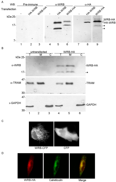
Fig. 2.
WRB is a 19 kDa membrane protein of the ER. (A) Identification of WRB–HA and HA–WRB. N-terminally or C-terminally HA-tagged forms of WRB (HA–WRB or WRB–HA) were expressed in HeLa cells. Proteins were separated by SDS-PAGE and analyzed by immunoblotting using the pre-immune serum (lanes 1–3), the anti-WRB serum (lanes 4–6) or anti-HA antibody (lanes 7–9). The asterisks indicate incomplete WRB. (B) Membrane association of WRB–HA. Untransfected HeLa cells (lanes 1–3) or HeLa cells transfected with HA–WRB (lanes 4–6) were analyzed by SDS-PAGE and immunoblotting using anti-WRB, anti-TRAM and anti-GAPDH antibodies. HeLa cells were permeabilized with digitonin, and cell constituents separated into a cytosolic and membrane fractions by centrifugation. Proteins of whole cell lysate (lanes 1 and 4), membrane (lanes 2 and 5) and cytosolic fractions (lanes 3 and 6) were analyzed. (C) Localization of WRB–CFP. CFP-tagged WRB (WRB–CFP) or CFP were expressed in RPE-1 cells and visualized by fluorescence microscopy. (D) ER localization of WRB–HA. WRB–HA was expressed in RPE-1 cells, immunostained with an anti-HA antibody and visualized with a fluorescent secondary antibody (red) by fluorescence microscopy. As an ER marker, calreticulin was visualized similarly using an anti-calreticulin antibody and a fluorescent secondary antibody (green).

