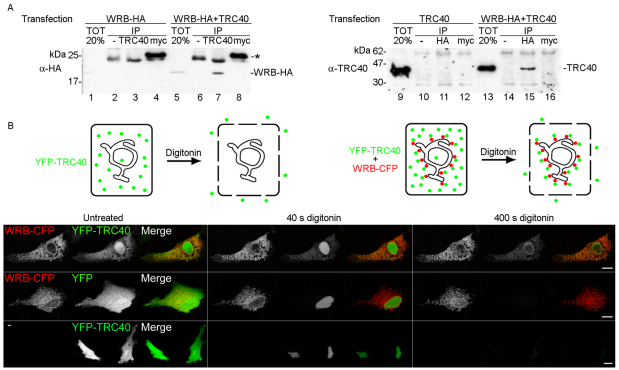
Fig. 3.
TRC40 associates with WRB–HA at the ER. (A) Co-immunoprecipitation of WRB–HA with TRC40 (left panel) and TRC40 with WRB–HA (right panel). HeLa cells were transfected with WRB–HA alone and with WRB–HA and TRC40 (left panel), or with TRC40 alone and with WRB–HA and TRC40 (right panel). 20% of the cell lysates were directly loaded onto the gel (lanes 1, 5 and 9, 13) and the rest were used for immunoprecipitation with the anti-TRC40 antibody (lanes 3 and 7), the anti-HA antibody (lanes 11, 15), an unrelated antibody (anti-myc) (lanes 4, 8 and 12, 16) or without an antibody (lanes 2, 6 and 10, 14). Lysates (TOT) and immunoprecipitates (IP) were analyzed by SDS-PAGE and immunoblotting using the anti-HA (left panel) or anti-TRC40 (right panel) antibodies. The asterisk indicates light chains of anti-myc antibody. (B) Colocalization of YFP–TRC40 with WRB–CFP at the ER. WRB–CFP was co-expressed with YFP–TRC40 (upper panel) or with YFP (middle panel), or YFP–TRC40 was expressed alone (lower panel). Cells were permeabilized with digitonin to release cytosolic content, and the CFP (red), YFP (green) signals or both signals were recorded over time using confocal microscopy. Untreated cells or cells treated with digitonin for 40 seconds and 400 seconds are shown. Scale bars: 10 μm. A graphical interpretation of the colocalization of YFP–TRC40 (green) with WRB–CFP (red) is shown on top of the figure.

