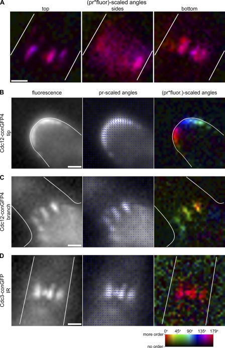
Figure 2.
Septin rings and multiple septin subunits are ordered throughout A. gossypii development. (A) A. gossypii septin rings analyzed at their top, middle, and bottom planes were measured to have GFP dipoles oriented perpendicular (or parallel, depending on the strain imaged) to the cell growth axis. The same septin ring shown in Fig. 1 (A and B), from a cell expressing Cdc12-conGFP4, is pictured here at different focal planes. The amount and azimuth of ordered septin protein is shown using a color scale (“pr*fluor-scaled angles” image). Cells expressing Cdc12-conGFP4 or Cdc3-conGFP from a replicating plasmid were imaged using polarized excitation and analyzed. Fluorescence (left), fluorescence overlayed with blue lines scaled in length by pr and oriented at the calculated GFP dipole angle (azimuth) for each camera pixel (middle), and the amount and azimuth of ordered septin protein using a color scale (right) are displayed. (B) Septins before ring assembly are ordered at growing hyphal tips. The typical azimuth orientation is perpendicular to the cell cortex. (C) Septin rings formed at branch points are highly ordered. The typical azimuth orientation is perpendicular to the cell growth axis. (D) An inter-region (IR) septin ring, assembled in the wake of a growing tip in a cell expressing Cdc3-conGFP. Septin rings in cells expressing this Cdc3-conGFP construct exhibit similar order, with the typical azimuth orientation perpendicular to the cell growth axis. Cell outlines are shown in white. Bars, 1 µm.

